Fairfield Medical Center Single Color Logo

Diagnostic Imaging

Chest Xray Review
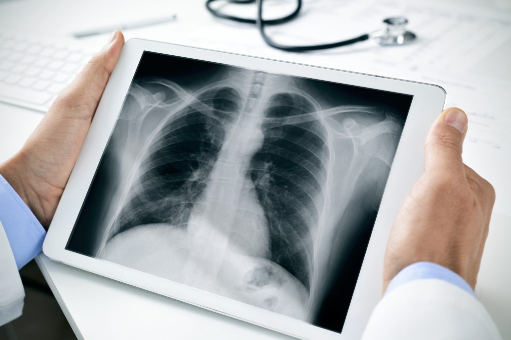
Imaging is an important tool for diagnosing and monitoring health conditions. Some types of imaging have overlapping purposes, and your provider will help determine which method is best for you.

Before arriving at a lab or imaging center, ask your doctor or technician for instructions on how to best prepare for your test. For example, some scans require a full bladder or an empty stomach. You should also inform your provider of any pre-existing health conditions, like pregnancy or implanted devices, and talk with them about any questions or concerns you may have.

In most cases, you will not receive results from your imaging at the time of your procedure. Instead, a technician or technologist will perform the procedure, and the images will be sent to your provider or a specialist for close review and interpretation. Once the images have been “read”, your provider will contact you with the results and next steps.


Understanding Imaging

Diagnostic imaging can give your provider a better picture of your health.

  • X-ray uses a specific type of radiation that passes through the body to create a picture of internal structures. X-rays are commonly used to evaluate bones for fractures or abnormalities but may also be used to diagnose some soft tissue and organ conditions.

    During the procedure, you will be positioned in either a standing, seated or lying position to place the area of concern in the imaging field. It’s important to note the dose of radiation you get from an x-ray is very small and poses no immediate threat to your health.

  • CT stands for computerized tomography scan. This minimally invasive technology uses radiation to capture images of the body at different angles. A computer then organizes the images to create a cross-sectional view of the body’s bones, blood vessels and soft tissues. CT scans provide greater detail than x-rays alone.

    During the procedure, you will lie on a table that moves through a scanning ring. You will be asked to remain still to keep images as clear as possible. Some tests may require contrast to help highlight areas of concern, and this medication may be given by mouth, rectally or by IV. If you are receiving a test that requires IV contrast, speak to your healthcare provider about any necessary blood work or screening before your appointment.

  • MRI stands for magnetic resonance imaging. This minimally invasive technology uses radio waves to create three-dimensional views of the body’s soft tissue. For example, an MRI may be used to assess damage to the muscles, joints, and ligaments (which are difficult to assess with x-ray) or take a closer look at internal organs, like the brain, heart and digestive tract.

    Before your imaging is completed, the technologist will review your medical history with you to ensure it is safe to have an MRI. During the procedure, you will lie on a table that moves into a large, tube-shaped machine. Depending on the exam, the technologist may place a “coil” over the area of interest. This device acts like an antenna, making the images clearer and well-defined.

    Much like CT scans, some tests may require contrast to help highlight areas of concern, and this medication may be given by given by mouth, rectally or by IV. If you are receiving a test that requires IV contrast, speak to your healthcare provider about any necessary blood work or screening before your appointment.


  • An ultrasound may also be called a sonogram. This procedure uses sound waves to create images of the structures within the body. While this technology is often associated with pregnancy, it can be used for a wide variety of purposes. Your provider may order an ultrasound to evaluate blood flow, explore reproductive health or diagnose conditions like gallbladder, thyroid or prostate disease. It may even be used to assess inflammation in the joints.

    During the procedure, a small hand-held probe (transducer) is most commonly pressed against the outside of the body. When the sound waves sent into the body encounter tissue, fluid or other internal structures, they bounce back and allow a computer to create an image of the examined area.

  • A mammogram is an x-ray picture of the breast that can be used for diagnostic or screening purposes. During the procedure, each breast is compressed to spread out the tissue, and an x-ray image is taken to look for abnormal changes.

    Fairfield Medical Center’s River Valley Campus offers a comfortable and serene space dedicated to Women’s Health. Learn more about what to expect during your mammogram.

  • Nuclear medicine is a special x-ray technology that can be used to evaluate organ function. For diagnostic imaging, a small amount of radioactive material, or tracer, will be given by mouth or as an injection. This medicine is absorbed by the body’s tissue. Next, a specially trained technologist will follow the tracer using a radiation detector to determine how it interacts with organs and tissues in the body.

    Your provider may order a nuclear medicine exam to assess kidney or thyroid function, identify areas of infection or inflammation throughout the body or learn more about a confirmed or suspected diagnosis.

  • Unlike other types of imaging, fluoroscopy uses live x-ray to create a moving picture of the body’s internal workings. In the outpatient setting, this is most commonly used for evaluation of the digestive tract, but it may also be used to assess the urinary tract or musculoskeletal system, like the spine or joints. This type of imaging requires the use of contrast, which may be administered differently depending on your condition.

    • Digestive (GI) exams – These tests highlight the stomach, small bowel or large bowel. During the exam, a radiologist will watch as the contrast moves through the digestive system to identify abnormalities or areas of concern. Depending on the area of study, you may be asked to drink the contrast or to receive an enema for rectal administration. Types of fluoroscopy GI exams include: barium enemas, upper GI exams, esophagrams and modified barium swallows.
    • Urinary exams – These tests evaluate the bladder, ureters and kidneys and may also be referred to as cystograms. During this procedure, a urinary catheter is used to administer the contrast for evaluation.
    • Musculoskeletal exams – If joint conditions cannot be diagnosed with standard imaging, fluoroscopy may be helpful. During an arthrogram, contrast is injected into the joint with a sterile needle. You may be asked to move the joint to allow for closer evaluation, or your provider may use this tool to assist with other treatments, like aspirations or injection. Additionally, a myelogram may be performed to evaluate the spine for injury, narrowing, nerve issues or growths. For this test, a sterile needle delivers contrast to the spinal column.

    As with any exam, there are variations of each test based on your individual health. If your provider orders fluoroscopy, be sure to speak with them about concerns, potential findings and next steps.

On entering the facility, I was greeted by the people at the desk and was able to help me get registered and gave helpful directions.”
- RVC Imaging Patient
I always go to this facility for all my tests mammogram, bone density scans, etc. I like the comfort, cleanliness and friendliness of this facility.”
- RVC Imaging Patient
Very quick service and friendly staff.”
- RVC X-Ray Patient
The lady who did my mammogram was very courteous and knew what she was doing. She made my experience very pleasant and quick.
- RVC Mammography Patient
The staff were excellent! I asked questions and I was answered thoroughly, and I was absolutely cared for in the best way! I was constantly asked if I was comfortable, and I felt very comfortable the entire procedure. Thank you for this!”
- RVC Mammography Patient

Preparing for Imaging

Being well-informed can help you feel confident and involved in your care.

If your provider recommends imaging, it’s helpful to ask these questions:

  • What kind of imaging will I be having?
  • What is the purpose of this test?
  • Are there any risks I should be aware of?
  • Will I receive any additional medication or injections for this test?
  • How can I best prepare for this test?
  • When can I expect to receive results?

If you are unsure about how to prepare for your test, call the office before your scheduled appointment and ask for clarity. Depending on your test, these questions may be useful:

  • Am I allowed to eat or drink before my test?
  • How should I take my prescribed medication the day of my test?
  • Are you aware that I have an implanted device?
    (Pacemaker, IUD, artificial joints or heart valve, bone or neuro stimulator, insulin pump, etc.)
  • Should I have a full or empty bladder?
  • Do I need to complete any screening labs or blood work before my test?

When arriving for your test, it is helpful to keep the following in mind:

  • Please have your ID and insurance card(s) available.
  • Please arrive for your test at least 15 minutes early.
  • Inform your provider if you have a history of allergies to contrast or dye.
  • Dress comfortably and leave valuables at home. You may be asked dress in a gown or to remove jewelry and accessories.

Accredited Care Gold Standard in Imaging

Fairfield Medical Center has been awarded accreditation in MRI, CT and mammography images by the American College of Radiology. The ACR gold seal of accreditation represents the highest level of image quality and patient safety.

When you receive diagnostic imaging services at FMC, you can rest assured that you are receiving care from a facility that prioritizes every aspect of care, including expertly trained personnel and advanced equipment in an inviting environment.

Imaging Locations

Fairfield Medical Center offers imaging services in several convenient locations. It’s important to note that less complex procedures, like x-rays, may be accommodated on a walk-in basis, but other tests may require scheduling and preparation. Additionally, certain tests may only be available at select locations. Your team will inform you of the most appropriate process.

Learn more about lab & imaging services available at FMC locations.